


- Download PDF
- |
- Download Citation
- |
- Email a Colleague
- |
- Share:
-
- Tweet
-
Journal of Biochemical & Microbial Technology
Volume 1, Issue 2, November 2013, Pages 16–19
Methodology Open Access
Sanfilippo syndrome: Cellulose affinity purification of α-N-acetylglucosaminidase for uptake studies
-
Truelson SN1 and
Choy FYM1,*

*Corresponding author: Francis Y. M. Choy, Department of Biology, Biomedical Research Centre, University of Victoria, 3800 Finnerty Road, Victoria, BC, Canada V8P 5C2. Telephone: (250) 721-7107; Fax: (250) 721-7120; E-mail: fchoy@uvic.ca
Received 26 July 2013 Revised 25 September 2013 Accepted 5 October 2013 Published 12 October 2013
DOI: http://dx.doi.org/10.14312/2053-2482.2013-3
Copyright: ©2013 Truelson SN, et al. Published by NobleResearch Publishers. This is an open-access article distributed under the terms of the Creative Commons Attribution License, which permits unrestricted use, distribution and reproduction in any medium, provided the original author and source are credited.
AbstractTop
Human α-N-acetylglucosaminidase (Naglu) is a lysosomal acid hydrolase deficient in Mucopolysaccharidosis III type B (Sanfilippo syndrome B). We have expressed Naglu in Spodoptera frugiperda 9 (Sf9) cells as a fusion protein with the cellulose binding affinity domain and an eleven amino acid protein transduction domain (PTD) from the HIV-Tat protein. Our aims are to develop an affinity method to purify Naglu efficiently and to facilitate delivery of Naglu across the blood-brain barrier, thus allowing enzyme replacement therapy to treat neurological symptoms. Cellulose affinity purification, followed by removal of the affinity tag via a Factor Xa cleavage site, allowed simple and efficient production of highly purified and catalytically active Naglu-PTD enzyme for uptake and cell permeation studies, with 382-fold purification and 18% yield from concentrated Sf9 culture medium.
Keywords: α-N-acetylglucosaminidase; lysosomal enzyme; cellulose affinity purification; Cex cellulose binding domain; Factor Xa; Sanfilippo syndrome; Sf9 cell expression system
IntroductionTop
Mucopolysaccharidosis III type B (MPS IIIB, Sanfilippo syndrome type B, OMIM #252920) is a lysosomal storage disorder caused by a deficiency in the enzyme α-N-acetylglucosaminidase (Naglu). Naglu catalyzes an intermediate step in heparan sulfate (HS) degradation by hydrolyzing the α-(1,4)-glycosidic bond between N-acetylglucosamine and uronic acid; inability to perform this step results in impaired degradation and build up of HS in lysosomes. Though somatic symptoms of MPS IIIB are relatively mild, the implications of HS buildup in the central nervous system are severe, resulting in neurodegeneration, behavioural problems, and shortened lifespan [1].
Several lysosomal storage disorders, including Mucopolysaccharidoses I, II, and VI, can be treated with enzyme replacement therapy. So far, enzyme replacement therapy has been unsuccessful in treating MPS IIIB due to the inability of intravenously-administered Naglu to cross the blood-brain barrier. The blood-brain barrier is an obstacle for many potential drug treatments that require central nervous system access, thus the discovery of various cell-penetrating peptides has provided hope for addressing this issue. In an attempt to facilitate the transport of therapeutic Naglu across the blood-brain barrier, our laboratory has created a cell-penetrating peptide-enzyme fusion with a modified version of the protein transduction domain (PTD) from the HIV-I transactivator of transcription (TAT) protein [2, 3].
The transduction of TAT PTD across biological membranes is believed to primarily occur through the endocytic process of macropinocytosis [4, 5]. Demonstration of the PTD’s ability to facilitate passage of fusion partners across membranes has resulted in investigation into TAT PTD-based therapies, such as for intracellular targeting of cancer cells [6-8]. Also, TAT PTD’s capacity to transport intravenously-administered fusion molecules across the cell membrane and blood-brain barrier in mice may result in revolutionary treatments for focal ischemia after stroke [9-11], mitochondrial disorders [12], and lysosomal storage diseases with visceral organ involvement and central nervous system involvement [13, 14].
This report describes a cellulose affinity method for purification of the Naglu-TAT PTD fusion enzyme (modTAT●Naglu) for potential enzyme replacement therapy. The construct, which was produced via secretory expression from Spodoptera frugiperda 9 (Sf9) cells, contained an N-terminal cellulose binding domain (CBD) from the Cellulomonas fimi exoglucanase Cex [15], followed by the Ile-Glu-Gly-Arg Factor Xa recognition sequence for subsequent seamless excision of the affinity tag. Cellulose affinity purification is an attractive purification method due to the low cost substrate and high specificity of the CBD for cellulose [16]. The described cellulose affinity protocol resulted in a 382-fold purification of active modTAT●Naglu with a yield of 18% from concentrated Sf9 culture medium.
Materials and methodsTop
Sf9 cultures with the stably integrated modTAT●Naglu expression plasmid (Figure 1) were prepared as described previously from our laboratory [2]. Large scale cultures were seeded and harvested according to the protocol of Vaags [17].

Figure 1 1 Sf9 plasmid vector constructs.
Abbreviations: ie2 (Orgyia pseudotsugata nucleopolyhedrovirus immediate early 2 promoter), TF (human transferrin secretion signal), CBD (cellulose binding domain from Cellulomonas fimi Cex), FX (Ile-Glu-Gly-Arg Factor Xa recognition sequence), modTAT●Naglu (Naglu fused to protein transduction domain of HIV-I transactivator of transcription protein), pA (immediate early 2 polyadenylation signal sequence), Zeo (Zeocin resistance gene).
Crude medium was concentrated 15-fold using an Amicon 8200 Stirred Ultrafiltration Cell (Millipore, Billerica, MA) at 25 psi pressure, 4oC with a 10,000 NMWL polyethersulfone membrane. Concentrated medium was then adjusted to 1 M NaCl, 0.3% Triton X-100, pH 9.0. After incubation on ice with gentle shaking for 1 h, the medium was centrifuged at 15,000 x g for 5 min. to remove precipitate. One mL of medium was added to 15 mg of Sigmacell Cellulose Type 101 (Sigma-Aldrich Canada, Oakville, ON) equilibrated with wash buffer (50 mM Tris-HCl, 1 M NaCl, 0.3% Triton X-100, pH 9.0) and incubated for 2 hrs on ice with vigorous shaking. The cellulose pellet was then washed with wash buffer, distilled water, and 1X Factor Xa Cleavage/Capture Buffer (EMD Biosciences, San Diego, CA). To prepare for Factor Xa digestion, the bulk of Factor Xa Cleavage/Capture Buffer was removed, and the cellulose was resuspended in the remaining buffer to form a thick slurry. Twelve units of Novagen Factor Xa (EMD Biosciences, San Diego, CA) were added to the slurry and incubated for 18-20 hrs at 37oC with vigorous shaking. Protein was then eluted from the cellulose in 100 µL increments and fractions with the highest Naglu activity were pooled (~first three). Factor Xa removal was performed using Xarrest Agarose (EMD Biosciences, San Diego, CA) according to the manufacturer’s instructions. All centrifugation steps were performed in a microcentrifuge at 15,000 x g for two min.
Naglu activity was measured using the 4-methylumbelliferyl-N-acetyl-α-D-glucosaminide fluorogenic substrate according to the protocol of Zhao and Neufeld [18]. Purity of samples was assessed using sodium dodecyl sulphate (SDS) polyacrylamide gel electrophoresis (PAGE) followed by protein staining using silver dye [19]. Protein concentrations were measured with the Bio-Rad Protein Assay kit (Bio-Rad, Hercules, CA) using bovine serum albumin as standard.
Results and discussionTop
Use of the Cellulomonas fimi Cex CBD allowed simple and efficient purification of active modTAT●Naglu enzyme that appears as a major protein band of ~80 kD (the expected size) upon SDS-PAGE and protein staining using silver dye (Figure 2, lane 3). The enzyme was purified 382-fold with a yield of 18% from concentrated Sf9 medium (Table 1). Yield from unconcentrated medium was closer to 13%, as use of the Amicon apparatus under described conditions typically resulted in 70% retention of modTAT●Naglu (data not shown). Attempts to concentrate modTAT●Naglu secreted in culture medium using ammonium sulfate precipitation was unsatisfactory, as ammonium sulfate apparently reacted with ingredient(s) in the SF900 medium that formed a colloidal-like buoyant substance, trapping and thus preventing modTAT●Naglu from recovery using centrifugation, even at 50,000 x g for more than one hr.
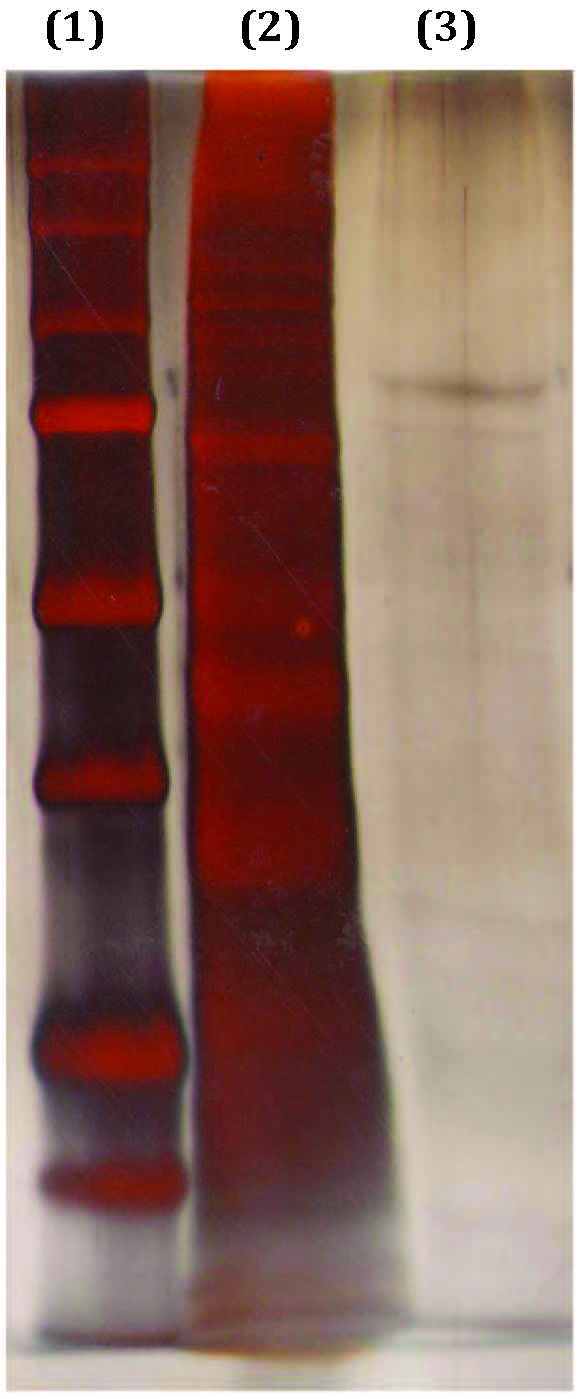
Figure 2 Silver stained SDS-PAGE gel indicating modTAT●Naglu enzyme following cellulose affinity purification. Lane 1: 5µL of protein standards (BioRad Precision Plus cat.#161-0374), M.W. (kD) from top: 250, 150, 100, 75, 50, 37,25, 20; lane 2: crude Naglu medium (6 µg of protein); lane 3: Naglu purified by cellulose affinity resin (35 ng of protein). Mobility of the Naglu protein band was between the 100 and 75 kD standard proteins and the size of the purified enzyme was estimated to be about 80 kD using standard protein markers in lane 1.
| Purification sample | Specific activity* (U per µg of protein) | Purification (fold) | Yield (%) |
| Crude concentrated medium | 0.034 | 1 | 100 |
| Post-cellulose affinity | 2.32 | 68 | 24 |
| Post-Factor Xa removal | 13 | 382 | 18 |
Binding specificity of Sigmacell Type 101 cellulose varied dramatically based on the condition of crude medium. Several conditions were tested, and binding specificity was assessed visually through direct denaturation of bound proteins from cellulose followed by SDS-PAGE and staining with silver dye. Binding with unaltered, concentrated medium provided little purification, since SDS-PAGE and silver dye staining of eluted Naglu showed that the protein bands were similar to that of the crude, unpurifed enzyme (Figure 2, lane 2), while alteration of medium to 0.1% Triton X-100, pH 8.5 provided a reduction in non-specific binding [2]. Increasing the ionic strength of crude medium resulted in a significant improvement, with the highest binding specificity achieved through alteration of medium to 1 M NaCl, 0.3% Triton X-100, pH 9.0. The on-cellulose Factor Xa digest to release modTAT●Naglu provided additional purification, as strongly absorbed contaminating proteins were removed with the cellulose pellet. It is of interest to note that while some CBD-fusion proteins can easily be eluted from cellulose using water [20], this is not the case for modTAT●Naglu as its affinity binding to cellulose is very strong and requires proteolytic cleavage using Factor Xa for recovery from the cellulose resins.
The specific activity of Naglu purified from CHO (Chinese hamster ovary) cells reported by Zhao and Neufeld at 30,000 units per mg protein [18] is higher than that of modTAT●Naglu as reported in this communication. This may be attributable to the difference in amino acid sequence between Naglu and modTAT●Naglu, the later being a chimeric enzyme fused with the eleven amino acid HIV-Tat protein transduction domain, as well as the difference between CHO and Sf9 cell expression systems.
Naglu is a lysosomal hydrolase with an optimal pH for activity of 4.5 [21]. Although the described protocol required a large increase in culture medium pH, there was no apparent loss of Naglu activity, as activity was restored upon returning crude medium to its original pH of around 5.8 (data not shown). To best preserve Naglu activity, most protocol steps were performed at refrigerating temperatures or on ice. Naglu, however, has been shown to remain stable at high temperatures, with the purified enzyme withstanding incubation for one hr at 50°C without significant loss of activity [21]. Though some Naglu may have been inactivated during the prolonged 37°C Factor Xa cleavage step, this condition resulted in optimal retrieval of active modTAT●Naglu.
ConclusionTop
We have expressed human recombinant Naglu as a catalytically active fusion enzyme with the eleven amino acid HIV-Tat protein transduction domain and a cellulose-binding affinity domain in cultured Sf9 cells. The recombinant enzyme secreted in the culture medium was adsorbed onto cellulose resins (Sigmacell Type 101) and purified 382-fold with 18% overall yield upon washing, recovery using Factor Xa digestion, and removal of the affinity tag using Xarrest Agarose (EMD Biosciences). The described protocol provides an effective cellulose affinity method for purification of Naglu. In addition, the low cost, availability, high binding specificity of cellulose under defined experimental conditions, and relative simplicity of this one-step affinity purification method will permit scaled-up enzyme production for transport and uptake studies for potential enzyme replacement therapy.
Acknowledgement
The authors thank Michael Chan for his invaluable technical assistance.
Funding
This work was supported by the Natural Sciences and Engineering Research Council of Canada Discovery Grant #138216-09 to F.Y.M.C. and a Pre-Doctoral Research Fellowship by the Canadian Society for Mucopolysaccharide and Related Diseases-Sanfilippo Children Research Foundation to S.N.T.
Conflict of interest
The authors wish to express that they have no conflict of interest.
ReferencesTop
[1] Valstar MJ, Bruggenwirth HT, Olmer R, Wevers RA, Verheijen FW, et al. (2010) Mucopolysaccharidosis type IIIB may predominantly present with an attenuated clinical phenotype. J Inherit Metab Dis 33:759–767. Article Pubmed
[2] Bandsmer JC, Campbell TN, Cheyne I, Choy FY (2006) Expression of active alpha-N-acetylglucosaminidase/TAT Chimerae in cultured Spodoptera frugiperda cells. Protein Pept Lett 13:353–356. Article Pubmed
[3] Ho A, Schwarze SR, Mermelstein SJ, Waksman G, Dowdy SF (2001) Synthetic protein transduction domains: enhanced transduction potential in vitro and in vivo. Cancer Res 61:474–477. Article Pubmed
[4] Gump JM, June RK, Dowdy SF (2010) Revised role of glycosaminoglycans in TAT protein transduction domain-mediated cellular transduction. J Biol Chem 285:1500–1507. Article Pubmed
[5] Nakase I, Takeuchi T, Tanaka G, Futaki S (2008) Methodological and cellular aspects that govern the internalization mechanisms of arginine-rich cell-penetrating peptides. Adv Drug Deliv Rev 60:598–607. Article Pubmed
[6] Ezhevsky SA, Nagahara H, Vocero-Akbani AM, Gius DR, Wei MC, et al. (1997) Hypo-phosphorylation of the retinoblastoma protein (pRb) by cyclin D:Cdk4/6 complexes results in active pRb. Proc Natl Acad Sci U S A 94:10699–10704. Article Pubmed
[7] Harbour JW, Worley L, Ma D, Cohen M (2002) Transducible peptide therapy for uveal melanoma and retinoblastoma. Arch Ophthalmol 120:1341–1346. Article Pubmed
[8] Zhao JF, Chen JY, Mi L, Wang PN, Peng Q (2011) Enhancement of intracellular delivery of anti-cancer drugs by the Tat peptide. Ultrastruct Pathol 35:119–123. Article Pubmed
[9] Cai B, Lin Y, Xue XH, Fang L, Wang N, et al. (2011) TAT-mediated delivery of neuroglobin protects against focal cerebral ischemia in mice. Exp Neurol 227:224–231. Article Pubmed
[10] Kilic E, Dietz GP, Hermann DM, Bähr M (2002) Intravenous TAT-Bcl-Xl is protective after middle cerebral artery occlusion in mice. Ann Neurol 52:617–622. Article Pubmed
[11] Kilic U, Kilic E, Dietz GP, Bähr M (2003) Intravenous TAT-GDNF is protective after focal cerebral ischemia in mice. Stroke 34:1304-1310. Article Pubmed
[12] Rapoport M, Salman L, Sabag O, Patel MS, Lorberboum-Galski H (2011) Successful TAT-mediated enzyme replacement therapy in a mouse model of mitochondrial E3 deficiency. J Mol Med (Berl) 89:161–170. Article Pubmed
[13] Higuchi K, Yoshimitsu M, Fan X, Guo X, Rasaiah VI, et al. (2010) Alpha-galactosidase A-Tat fusion enhances storage reduction in hearts and kidneys of Fabry mice. Mol Med 16:216–221. Article Pubmed
[14] Schwarze SR, Ho A, Vocero-Akbani A, Dowdy SF (1999) In vivo protein transduction: delivery of a biologically active protein into the mouse. Science 285:1569–1572. Article Pubmed
[15] Pfeifer TA, Guarna MM, Kwan EM, Lesnicki G, Theilmann DA, et al. (2001) Expression analysis of a modified factor X in stably transformed insect cell lines. Protein Expr Purif 23:233-241. Article Pubmed
[16] McLean BW, Bray MR, Boraston AB, Gilkes NR, Haynes CA , et al. (2000) Analysis of binding of the family 2a carbohydrate-binding module from Cellulomonas fimi xylanase 10A to cellulose: specificity and identification of functionally important amino acid residues. Protein Eng 13:801-809. Article Pubmed
[17] Vaags A (2004) Expression and Purification of HIV-I TAT Protein Transduction Domain Fused with Acid β-glucosidase and Enhanced Green Fluorescent Protein. MSc Thesis, University of Victoria: Victoria, BC, Canada.
[18] Zhao KW, Neufeld EF (2000) Purification and characterization of recombinant human alpha-N-acetylglucosaminidase secreted by Chinese hamster ovary cells. Protein Expr Purif 19:202-211. Article Pubmed
[19] Mortz E, Krogh TN, Vorum H, Görg A (2001) Improved silver staining protocols for high sensitivity protein identification using matrix-assisted laser desorption/ionization-time of flight analysis. Proteomics 11:1359-1363. Article Pubmed
[20] Sugimoto N, Igarashi K, Samejima M (2012) Cellulose affinity purification of fusion proteins tagged with fungal family 1 cellulose-binding domain. Protein Expr Purif 82:290-296. Article Pubmed
[21] Sasaki T, Sukegawa K, Masue M, Fukuda S, Tomatsu S, et al. (1991) Purification and partial characterization of alpha-N-acetylglucosaminidase from human liver. J Biochem 110:842-846. Article Pubmed
 Copyright
© 2012-2025 NobleResearch Group. All Rights Reserved
Copyright
© 2012-2025 NobleResearch Group. All Rights Reserved